
Intraoperative laparoscopic view of the Phrygian cap gallbladder,... Download Scientific Diagram
What is it? A congenital abnormality of the gallbladder. Incidence: Found in 4% of the population. Appearance on Imaging: Can mimic a liver mass, leading to potential misinterpretation. Pathological Significance: None. It's not a disease or a harmful condition. Symptoms: Typically none.
Gallbladder Surgeon in Katy, TX Dr. Clay Albrecht Dr. Clay Albrecht
A Phrygian cap is a congenital abnormality of the gallbladder [ 4,5] and has an incidence of 4% [ 6 ]. It is the most common congenital anomaly of the gallbladder and can simulate a mass in the liver during hepatobiliary imaging [ 5 ], which may suggest a tumour. It can also simulate a duplication of the gallbladder [ 7 ].

PHRYGIAN CAP GALLBLADDER (OCT.2021) Dr.Raad Alsaffar
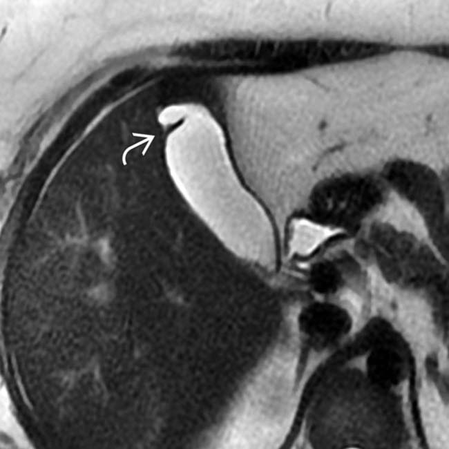
Phrygian cap is the most common congenital variant of gallbladder anatomy, with an incidence of 1-6% [1,2,3].It represents folding of the fundus of the gallbladder upon its body (Fig. 1) and resembles the soft conical cap worn by people of ancient Phrygia (central Turkey) (Fig. 2).Folding of the fundus during embryological development causes a Phrygian cap [].
The Gallbladder Radiology Key
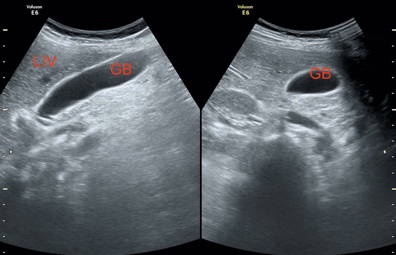
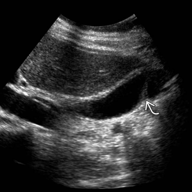
Phrygian caps are the most common congenital anatomic variant of the gallbladder . It denotes folding of the fundus back upon the gallbladder body and is asymptomatic with no pathological significance. Radiographic findings A Phrygian cap may be identified on ultrasound, multiphase CT/MRI, or cholescintigraphy 3 . Ultrasound

Ultrasound and Color Doppler videos Normal Gallbladder variants Phrygian cap
The normal gallbladder may exhibit folds, including the common and asymptomatic folding of the gallbladder fundus termed a "phrygian cap." Less commonly seen are true septa that can lead to stasis and stone formation. Anomalies in the number of gallbladders— ranging from absent to duplications—are rare but have also been reported [ 1 ]. Fig. 1A.

FongHealth Phrygian Cap Gall Bladder
Adenomyomatosis of the gallbladder is a hyperplastic cholecystosis of the gallbladder wall. It is a relatively common and benign cause of diffuse or focal gallbladder wall thickening , most easily seen on ultrasound and MRI. Epidemiology Adenomyomatosis is relatively common, found in ~9% of all cholecystectomy specimens 6.

Softtissue images. “Phrygian cap” gallbladder CJS
Phrygian cap is the most common of the gallbladder anomalies reported to be present in up to 18% of patients with a functioning gallbladder (Figs. 79.11 and 79.12) (Boyden 1935). An infolding of a septum between the body and the fundus creates the Phrygian cap deformity.

Phrygian cap Gallbladder ultrasound video YouTube
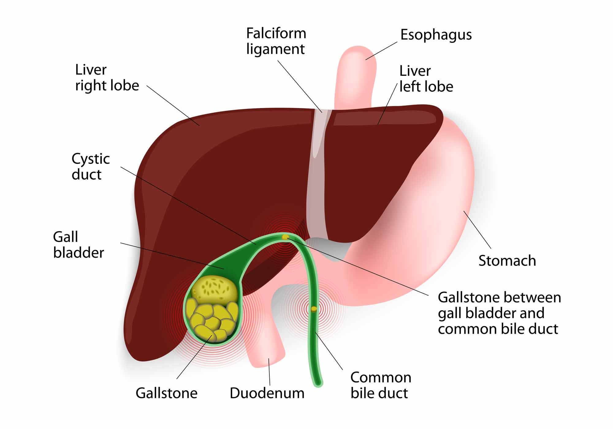
This is called a Phrygian cap and has no pathologic or surgical significance.[1] The gallbladder is a pear-shaped organ located in the right upper quadrant of the abdomen. It measures approximately 7 cm to 10 cm in length and 4 cm in width.
Gallbladder Radiology Key
In medicine, a Phrygian cap is the folded portion of some gallbladders that resembles the Phrygian cap (a soft conical cap with the top pulled forward, associated in antiquity with the inhabitants of Phrygia, a region of central Anatolia ). It is a normal anatomical variant seen in 1-6% of patients. [1]
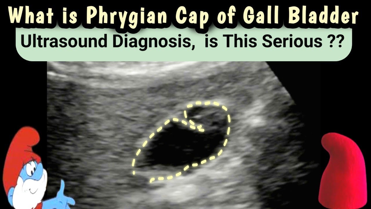
What is Phrygian Cap of Gall bladder Ultrasound Diagnosis is this serious YouTube
A Phrygian cap is a congenital anomaly of the gallbladder with an incidence of 4%. It can simulate a mass in the liver during hepatobiliary imaging and is sometimes mistaken for pathology. A Phrygian cap, however, has no pathological significance and normally causes no symptoms.
Congenital Abnormalities of the Gallbladder Radiology Key
A series of five fetuses with a Phrygian cap gallbladder, a condition infrequently reported in the antenatal period, is reported. In all cases, examination of the fetal gallbladder displayed the characteristic folding of the fundus over the body. No associated findings were detected. The gallbladder length was longer than normal in all cases.
Congenital Abnormalities of the Gallbladder Radiology Key
Adenomyomatosis is a benign condition that is pathologically characterized by hyperplasia of the gallbladder wall mucosa and muscularis propria, with pathognomonic epithelial invaginations forming cystic pockets (Rokitansky-Aschoff sinuses). These sinuses may contain calculi or cholesterol crystals.

Intraoperative laparoscopic view of the Phrygian cap gallbladder,... Download Scientific Diagram
Phrygian cap of the gallbladder is a triangular deformity of the gallbladder fundus and a fairly common anatomical variant.

Phrygian cap gallbladder Image
Phrygian cap: gallbladder fundus folding back upon the body resembling the head garment worn by inhabitants of Phrygia (modern Türkiye) 1200-700 BCE 1,2 Hartmann pouch: fold or diverticulum near the neck of the gallbladder where gallstones can often collect ref Radiographic features Ultrasound

Phrygian cap cholecystitis Surgery
The most common congenital anomaly of the biliary tract is a folded gallbladder. 1 Boyden described this anomaly as a "Phrygian cap" deformity in 1935 because it resembled a bonnet worn by the ancient Phrygians, 2 who lived in Asia Minor during the 12th century BC. 3 This type of gallbladder is thought to empty at a normal rate and, in contrast.

Phrygian cap illustration Radiology Case Medical illustration, Radiology
The Phrygian Cap Gallbladder is a congenital abnormality where the fundus, or the top portion of the gallbladder, folds over the body of the organ, forming a cap-like structure. This gives the gallbladder a distinctive appearance, resembling the shape of a Phrygian cap. How it Differs from a Normal Gallbladder Structure